

About 80% of breast cancer biopsies turn out benign – new imaging tool promises clearer diagnoses and fewer biopsies
- Written by Quing Zhu, Professor of Engineering, Washington University in St. Louis
Ultrasound is widely used[1] in breast cancer diagnosis. While it can effectively show that a lump is filled with fluid – indicating it is unlikely to be cancer – it cannot reliably determine whether a solid mass is benign or cancerous. This often leads doctors to order breast biopsies to confirm the presence of cancer.
However, most breast biopsies do not detect cancer. In the U.S., more than 1 million breast biopsies are performed each year, and about 80% of them are benign[2]. Unnecessary biopsies are linked to potential harms, including increased anxiety[3], complications from the procedure[4] and medical costs[5]. Despite advances in breast imaging, breast biopsy remains the only definitive method to determine whether a suspicious lump is cancerous.
My work as an engineer[6] focuses on improving imaging technology to detect and diagnose cancer. Breast cancer grows when the tumors form new blood vessels[7] and consume more oxygen. This makes examining blood vessels and oxygen levels potential biomarkers that could improve breast cancer diagnosis.
Diffuse optical tomography, or DOT[8], is an imaging technology that uses near-infrared light to measure total blood hemoglobin concentration and oxygen levels – key indicators of tumor activity – in the breast lump. It does not require patients to be injected with contrast dyes to make the image clearer.
My team and I found that combining ultrasound with DOT can improve the accuracy of breast cancer diagnoses[9] and reduce unnecessary breast biopsies. The ultrasound provides information about the structure of a breast lump, while DOT provides information about its function, and this data together can improve breast cancer diagnosis.
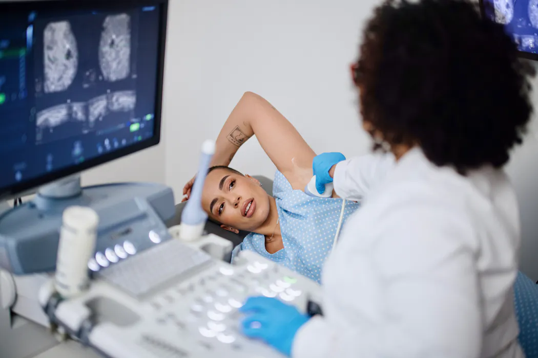
Anyone with breast tissue is at risk of developing breast cancer.Improving breast ultrasounds with DOT
In our study, we imaged 226 patients recommended for routine breast biopsy using our new hand-held imaging technology, which combines ultrasound with diffuse optical tomography. These patients had either breast cancer or benign lumps, and their final diagnosis was confirmed with a biopsy.
Radiologists initially evaluated each patient using standard imaging methods, such as ultrasound and mammography. They then reviewed additional information from DOT images. Importantly, the radiologists and engineers were blinded to the biopsy results when determining diagnoses.
We observed significant biological differences between cancerous and benign lumps[10]. Cancerous lesions had significantly higher levels of hemoglobin and lower levels of oxygen than noncancerous tissue. More aggressive cancers showed even higher hemoglobin concentrations and lower oxygen levels than less aggressive tumors.
When radiologists were able to review DOT measurements, biopsies of benign lumps decreased by approximately 25%[11]. The false-negative rate was 1.8%, which aligns with medical guidelines[12] that recommend monitoring rather than an immediate biopsy.
Future of breast cancer screening and diagnosis
Breast cancer is the most commonly diagnosed cancer in women[13] worldwide. There were approximately 2.3 million new cases and 670,000 deaths[14] reported in 2022. If these rates continue, researchers project around 1.1 million breast cancer-related deaths will occur in 2050.
More accurate, noninvasive diagnostic tools can not only reduce unnecessary biopsies but also lead to more precise and efficient diagnoses. Beyond ultrasound, researchers have also explored combining other imaging techniques with DOT, including X-ray mammography, 3D mammography[15] and MRI[16]. However, DOT systems combined with mammography and MRI are more difficult for routine use in the clinic[17] compared to ultrasound. My team is working to further refine our technology, including incorporating AI tools to help process imaging data.
Minimizing avoidable procedures can help preserve a patient’s quality of life and reduce health care costs. I believe these improvements can collectively have a meaningful and far-reaching effect on patient care and the broader health care system.
References
- ^ Ultrasound is widely used (www.cancer.org)
- ^ 80% of them are benign (www.bcrf.org)
- ^ increased anxiety (doi.org)
- ^ complications from the procedure (doi.org)
- ^ medical costs (doi.org)
- ^ as an engineer (scholar.google.com)
- ^ form new blood vessels (doi.org)
- ^ Diffuse optical tomography, or DOT (doi.org)
- ^ improve the accuracy of breast cancer diagnoses (doi.org)
- ^ differences between cancerous and benign lumps (doi.org)
- ^ decreased by approximately 25% (doi.org)
- ^ medical guidelines (my.clevelandclinic.org)
- ^ most commonly diagnosed cancer in women (doi.org)
- ^ 2.3 million new cases and 670,000 deaths (doi.org)
- ^ X-ray mammography, 3D mammography (doi.org)
- ^ and MRI (doi.org)
- ^ routine use in the clinic (doi.org)
Authors: Quing Zhu, Professor of Engineering, Washington University in St. Louis